|
Fifth Dimension Site Map Search Contact Us |
|||

|
Osteoporosis - Evaluation, Prevention and Therapy |
||
|
|||
Optimal Evaluation and Therapy
Back to the Table Of Contents
Criteria for diagnosis
The diagnosis of osteoporosis is based on bone mass measurements to establish risk of fracture. History and physical examination and laboratory assessments are necessary to establish the cause and to initiate appropriate therapy. When secondary causes are found, treatment for the cause as well as for the osteoporotic process is required. BMD correlates with capacity for load bearing. Accurate measurements of the site of interest, e.g. measurements of the femoral neck by DXA (dual-energy X-ray absorptiometry) to assess risk for hip fracture are more reliable than peripheral measurement, e.g., sonography of the knee or heel. T score is defined as the number of SD's (standard deviations) above or below the average BMD (bone mineral density) of a young healthy white woman. Osteoporosis is defined as -2.5 SD's. Osteopenia is defined as -1.5 to -2,5 SD (see above). Bone remodeling can be assessed by measurements of markers of bone turnover, such as serum bone specific alkaline phosphatase, osteocalcin, and urinary pyridinoline and deoxypyridinoline levels. These markers do not predict bone mass or fracture risk but do correlate with bone remodeling. From the practical point, serial bone density measurements, e.g. at intervals of 1-3 years, are the mast readily available means for evaluating diagnosis and treatment of osteoporosis.
Medical Treatment
Back to the Table Of Contents
Calcium and vitamin D intake modulates age-related increase in parathyroid gland hormone (PTH) levels and bone resorption. They can reduce fractures by maintaining BMD. The recommended dose of Vitamin D is 400-1000 IU /day. To achieve this optimum intake a combination of dietary calcium sources (See Fig. 6.6) and calcium in pill form (Fig 7) is usually needed. Calcium carbonate and calcium citrate preparations are preferred. In patients with mild acidosis, potassium bicarbonate may stabilize calcium balance.
Physical activity is essential for bone growth and maintenance. It also improves muscle strength and balance, thus decreasing the risks of fracture and falls. Low impact exercises such as walking 45 minutes/day have beneficial effects. The greatest benefit from exercise is in patients who had been inactive. Excessive exercise, especially in young women, associated with weight loss and amenorrhea can be detrimental to bone (e.g., anorexia nervosa).
Bisphonsphonates: alendronate (fosamax), etidronate(didronel), and residronate(actonel) can reduce risk of vertebral fracture by 30-59% as well as of non-vertebral fractures. The most commonly used bisphosphonate in the USA is fosamax. It is now available as a single oral dose of 70 mg given once/week with water. This has lessened the gastrointestinal side effects of the bisphosphonates. In severely debilitated patients who cannot take oral medication, e.g. transplant patients on steroids, injectable forms of bisphosphonates (Aredia and zometa) are available. The most potent and long-acting compound, zometa, given parenterally once or twice a year has become available recently.
In breast cancer patients who cannot be placed on estrogen, and who show osteopenia, the use of two non- estrogenic compounds has been shown to prevent progression of the disease process. The bisphosphonates Fosomax and Actonel, taken once weekly will halt or slow down the progressive bone loss, and may even protect the bones agaisnt attack by cancer cells. The SERM ( selective estrogen receptor modulator ) Evista, likewise ,has been approved by the FDA for the prevention of osteoporosis in patients who cannot take estrogen or bisphosphonates. Furthermore, Tamoxifen has a protective effect against breast cancer.
Too much sodium can also cause excessive losses of calcium in the urine. Diuretic medicines, which reduce calcium losses through the kidneys (Thiazides), preserve bone mass. Other diuretics (e.g. furosemide) may cause excessive calcium losses.
| Calcium Preparations Available (listed by type and calcium content; derived from various sources; generic preparations may be cheaper) |
|
| Type and Name | Elemental calcium per tablet (in mgs) |
| Calcium Carbonate Preparations | |
| Tums and Tums E-X | 200 or 300 |
| Alka-Mints | 340 |
| Bio-CaI | 250 or 500 |
| CalSup | 300 |
| Caltrate | 600 |
| Os Cal 500 | 500 |
| Titralac tablets | 168 |
| Titralac liquid (1 teaspoon) | 400 |
| Calcimax | 325 and 650 |
| Dicarbosil | 200 |
| Calcium Carbonate with Vitamin D | |
| OsCal and generic oyster shell calcium with Vitamin D | 250 plus 125 U Vitamin D |
| Dical-D | |
| capsule | 115 plus 133 U Vitamin D |
| wafers | 230 plus 200 U Vitamin D |
| Calel-D | 500 plus 200 U Vitamin D |
| Calcium Phosphate Preparations | |
| Dibasic Calcium phosphate | 112 |
| Posture | 600 |
| Calcium Citrate | |
| Citracal | 200 |
| "Chelated" Calcium Tablets | |
| Chelated calcium | 160 |
| Calcium Orotate | 50-100 |
| Calcium Lactate Tablets | 100 |
| Calcium Gluconate Tablets | 40-60 |
|
All calcium tablets are better absorbed if chewed before swallowing. |
|

Dairy and Non-dairy Sources of Calcium
| Food | Serving Size | Calcium (mg.) |
| Dairy | ||
| Milk | ||
| evaporated | 1 Can | 1034 |
| skin/non fat | 1 Can | 298 |
| low-fat | 1 Can | 297 |
| buttermilk | 1 Can | 296 |
| whole | 1 Can | 288 |
| chocolate | 1 Can | 415 |
| Yogurt, low-fat/non fat | 1 Can | 415 |
| Cheese | ||
| Parmesan | 1 oz. | 390 |
| Ricotta, part-skim | 1/2 cup | 337 |
| Swiss | 1 oz. | 272 |
| Cheddar | 1 oz. | 211 |
| Muenster | 1 oz. | 203 |
| Mozzarella | 1 oz. | 183 |
| American | 1 oz. | 174 |
| Camenbert | 1 oz. | 110 |
| Cottage cheese, low-fat | 4 oz. | 77 |
| Brie | 1 oz. | 52 |
| Non-Dairy | ||
| Fish | ||
| Sardines, with bones | 3 ounces | 372 |
| Salmon, with bones | 3 ounces | 167 |
| Oysters | 3 ounces | 113 |
| Shrimp, canned | 3 ounces | 98 |
| Vegetables (cooked): | ||
| Colard greens | 1 cup | 289 |
| Bok choy | 1 cup | 250 |
| Kale | 1 cup | 206 |
| Spinach* | 1 cup | 200 |
| Mustard greens | 1 cup | 193 |
| Broccoli | 1 med stalk | 158 |
| Muffins: | ||
| Bran | 1 medium | 142 |
| Corn | 1 medium | 105 |
| Almonds, whole | 1/2 cup | 166 |
| Tofu, processed with calcium sufate | 4 ounces | 145 |
| Blackstrap molsasses | 1 T | 137 |
| Tortilla, corn or flour, unrefined | 2 6" | 120 |
| Orange | 1 medium | 54 |
| *contains substances that block calcium absorption | ||
Hormonal replacement therapy (HRT) has been the mainstay of therapy over the years for the prevention and treatment of osteoporosis. The potential side effects, and especially the fear of breast and uterine cancer has made compliance with long-term use difficult. SERMs (selective serum estrogen-receptor modulators), such as raloxifene, may substitute for HRT and can reduce vertebral fracture risk by 36%.(as well as the risk of breast and uterine cancer) The role of tamoxifen in osteoporosis is unclear, although it appears to have a positive effect on reducing osteoporosis, but the incidence of uterine cancer is enhanced(this may be offset by the use of progesterone). The other SERM, Raloxifene (Evista), may become the treatment of choice for osteoporosis in patients who had breast and/or uterine cancer.
Phytoestrogens are weak estrogens. Thus far, they have no proven therapeutic effect on osteoporosis although they may help to reduce hot flushes. The cholesterol-lowering agents, the statins. (e.g. lovostatin) have been shown to have a mild bone-sparing effect but in the presently used doses they are not effective treatment for osteoporosis.
Salmon calcitonin injections, and especially nasal calcitonin may play a role in preventing and possibly in treating osteoporosis. Compliance is a problem, as there is a 60% drop out rate in the use of salmon calcitonin. It may have an analgesic effect in recent vertebral fractures.
Combination treatment schedules, e.g. combining HRT with a bisphosphonate are under investigation.
In severe osteoporosis, especially in elderly patients who are 3-5SD below normal, the experimental treatment with daily injections of a parathyroid hormone fraction (Forteo) has been associated with marked increases in bone density and cessation of fractures. The drug, which is costly, will be released soon.
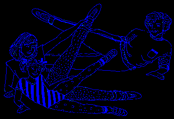
Aside from medical treatment, orthopedic measures, i.e. adequate but not excessive back support, physiotherapy, exercise programs (Figure 9), prevention of blood clots during periods of immobilization etc. are helpful adjuvant measures. In severely kyphotic patients, vertebroplasty involving injection of polymethylacrylate bone cement into the fractured vertebra may be helpful, but must still be considered an experimental procedure.
Exercise-The Second Key to Prevention
Back to the Table Of Contents
During all periods of life, especially childhood and the childbearing years, not only adequate diet but also exercise is necessary to help increase and maintain bone mass. Sporadic exercise won't do it. A regular program emphasizing weight bearing is essential. Carefully planned exercise programs are important, even for people confined to wheelchairs or bed for more than a few weeks, since extended sedentary periods can be themselves accelerate the development of osteoporosis. Exercise not only promotes overall well being but yields advantages specifically related to bone strength.
Certain exercises stimulate bones, promoting bone growth and strength. The best exercises for this purpose are walking, cycling, tennis, gymnastics, basketball, football, and soccer, but some of these sports present participation difficulties for many of us. Jogging in moderation is helpful, but excessive running can lead to cessation of regular menstruation due to a fall in estrogen and subsequent bone loss. Ballet dancers who severely limit their calories but participate in strenuous workouts have been known to suffer from this problem. These women are functionally in a premature menopause. Excessive weight loss, like in anorexia nervosa, can have the same effect and may be a major factor in causing exercise-induced osteoporosis. Swimming is a great exercise for cardiovascular fitness but less so for promoting bone strength, since it puts less stress on the bones. However, it helps mobility of joints and strengthens muscles.
Exercise generates tiny electrical currents throughout the bones that stimulate bone growth and remodeling. Exercise alters hormonal balances, favoring the hormones that protect bone, so long as the exercise is not excessive. Overall, an active lifestyle is most important in reducing the risk of osteoporosis. So walk rather than ride, climb stairs rather than take an elevator, and stand rather than sit wherever appropriate.
Elderly patients who have beginning osteoporosis should be protected against falling, especially when exercising. They need exercise adapted to their physical limitations.
Many patients with advanced osteoporosis require a lightweight back support, which is high enough to support both the lower and upper spine. It must not be too rigid but should be adequate to support the upright posture. A cane may be needed for stability. Padding of hips may prevent hip fracture.

The Importance of Good Posture
Back to the Table Of Contents
 Proper instruction in ways to sit, stand, bend and lift objects is greatly needed.
 The Proper Way to Sit. Support your lower back with a pillow or by a straight high-backed chair. When driving or reading, avoid bending the neck forward. When rising from a chair, do it slowly.
 The Proper Way to Walk and Stand. Keep your head high, look forward with the chin in. Pull your shoulders back, pull your stomach in to maintain the natural arch of the lower back, and tighten your buttocks. Wear low-heeled shoes with rubber soles. Wear a heel lift if necessary to balance the hips. Keep your toes pointing forward.
 The Proper Way to Lift.You must bend your knees when lifting heavy objects to avoid backstrain and further compression fractures. Use your Leg muscles rather than your back!
What are the Directions for the Future
Back to the Table Of Contents
There has been an explosion of interest in osteoporosis as a major worldwide public health problem. A great variety of options for the diagnosis and treatment have become available. The public has become informed about osteoporosis through the media, and osteoporosis has become a household word. Yet we are not certain about the best way to diagnose and to treat this multifactorial disorder.
Problems remain in the field of diagnosis. When and how should we start screening for bone loss? Ideally the first measurements should be done in the adolescent period to see if the maximum peak bone mass has been achieved, and preventive measures initiated at that time and maintained throughout life will have the greatest chance to achieve and maintain bone health. How should this be financed? Osteoporosis is largely a silent disorder, and prior to a clinical event, i.e. fracture, the patient may be unaware of having low bone mass. Should we screen only patients with known risk factors, i.e. family history of osteoporosis, postmenopausal status, age, poor nutrition etc. Which single measurement will be accurate and cost-effective?
The greatest problem in a largely asymptomatic disorder is compliance. It has been estimated that if a physician prescribes an effective treatment, e.g. estrogen (HRT), at the end of a year only some 20% of women continue to refill their medication. Serial bone density measurements have not markedly increased patient compliance, although it is a step in right direction, if it can be combined with an accurate and simple measurement of bone turnover.
We need a reliable validated fracture risk assessment tool that combines measurement of bone density with bone quality and strength. Since the majority of therapeutic agents currently available are largely antiresorptive, i.e. decreasing bone breakdown, search for simple, effective, and safe anabolic agents to restore lost bone mass is needed. The recently introduced intermittent injection of a recombinant parathyroid hormone fraction (Forteo), which has shown impressive increases and even normalization of lost bone mass, is a step in the right direction, and will probably be available soon. Other bone growth factors are under investigation.
The future looks bright. Multiple osteoporosis screening centers have appeared even in rural areas. The widespread shortcuts by cheap and simple bone screening devices located in drug stores and shopping centers must carry a caveat. They may over diagnose some patients who are needlessly frightened and subjected to unnecessary treatment, but worse yet, they may miss patients with established osteoporosis at regions not measured, e.g., the hip, and give the patients false security and delay in getting adequate treatment.
|
You are welcome to share this © article with friends, but do not forget to include the author name and web address. Permission needed to use articles on commercial and non commercial websites. Thank you. |